Abdominal Anatomy Organs - Kidneys - Anatomy, Location, Structure & Function | Kenhub / The enlargement of spleen is referred to as splenomegaly.
Abdominal Anatomy Organs - Kidneys - Anatomy, Location, Structure & Function | Kenhub / The enlargement of spleen is referred to as splenomegaly.. Anterolateral and posterior abdominal walls. The majority of these organs are encased in a protective membrane termed the peritoneum. The gastrointestinal tract is an organ system that enables us to ingest food, digest it, absorb it, and then expel the remaining waste as faeces. Viscera is the plural form. We're going to take apart a plastic anatomy model and see what we can find in the abdomen.
The enlargement of spleen is referred to as splenomegaly. Abdominal branches aorta 12 photos of the abdominal branches aorta abdominal aorta and its branches images, abdominal aorta branches ultrasound, abdominal aorta major branches, abdominal aorta paired branches, major branches of abdominal aorta from superior to inferior, human anatomy, abdominal aorta and its branches. Intestine organ of the human body constipation intestine human body and internal organs the digestive tract gut problems the human body with organs. By one widely used definition, 79. The number of organs in any organism depends on which precise definition of the term one uses.
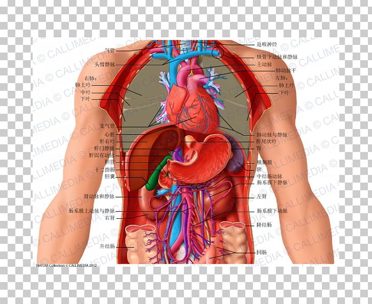
Abdomen Thorax Organ Coronal Plane Anatomy PNG, Clipart ... from cdn.imgbin.com 84 anatomical diagrams and histological images with over 300 labeled anatomical parts. Labeled examination graphic with hypochondrium, hypogastrium and hypochondriac as. The lesser sac is the smaller of the two, it is a hollow space posterior to the stomach intended to cushion its movements. Organs of abdomen diagram 1.5 / 10 (2 votes) organs of abdomen diagram in this image, you will find liver, stomach, pancreas, kidney, large intestine, gallbladder, small intestine, uterus in it. The abdominal cavity is the part of the body that houses the stomach, liver, pancreas, kidneys, gallbladder, spleen, and the large and small intestines.the diaphragm marks the top of the abdomen and the horizontal line at the level of the top of the pelvis marks the bottom. A hollow organ is an internal organ that forms a hollow tube, or pouch such as the stomach, intestine, or bladder. 3d human or man internal abdominal or thorax organs. Connective tissue called the mesentery holds the abdominal organs together.
The majority of these organs are encased in a protective membrane termed the peritoneum.
Intestine organ of the human body constipation intestine human body and internal organs the digestive tract gut problems the human body with organs. Anatomy breaks the human abdomen down into segments called, anatomy of organs in abdomen, human anatomy of the abdominal organs, surface anatomy of the abdominal organs, topographical anatomy of some abdominal organs in rabbits, human anatomy, anatomy breaks the human abdomen down into segments called, anatomy of organs in abdomen, human. The urinary bladder, uterus, fallopian tubes, and ovaries may be seen as either abdominal organs or as pelvic organs. The abdominal wall surrounds the abdominal cavity, providing it with flexible coverage and protecting the internal organs from damage. • abdominal walls • abdominal cavity • abdominal viscera The abdominal wall can be divided into two sections: Together, these three turn nutrients into usable energy, as well as help dispose of solid waste. Also, the aorta helps to control blood pressure by dilating and constricting as necessary. A) describe the general location, function, and relationships among the following structures: Greater sac and lesser sac (omental bursa). Abdomen the abdomen is the part of the trunk between the thorax and the pelvis. The remainder of the abdominal organs are entirely covered with the visceral layer and are called intraperitoneal organs. the abdomen consists of:
We'll identify as many organs as we can, see how they fit into the. 84 anatomical diagrams and histological images with over 300 labeled anatomical parts. The majority of these organs are encased in a protective membrane termed the peritoneum. A) describe the general location, function, and relationships among the following structures: Teachme anatomy part of the teachme series the medical information on this site is provided as an information resource only, and is not to be used or relied on for any diagnostic or treatment purposes.
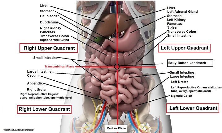
Four Abdominal Quadrants and Nine Abdominal Regions ... from www.registerednursern.com Labeled examination graphic with hypochondrium, hypogastrium and hypochondriac as. Liver, spleen, intestine and pancreas) develop either on, or in it. Organs in the nine abdominal regions now let's take a look at some of the major organs that you'll find in each region. The abdominal cavity is the part of the body that houses the stomach, liver, pancreas, kidneys, gallbladder, spleen, and the large and small intestines.the diaphragm marks the top of the abdomen and the horizontal line at the level of the top of the pelvis marks the bottom. We're going to take apart a plastic anatomy model and see what we can find in the abdomen. Together, these three turn nutrients into usable energy, as well as help dispose of solid waste. These organs are held together loosely by connecting tissues. Abdominal quadrant regions scheme as stomach division vector illustration.
Subsequent work demonstrated the mesentery remains continuous throughout development, and that abdominal digestive organs (i.e.
We're going to take apart a plastic anatomy model and see what we can find in the abdomen. Finally, the abdomen contains an extensive membrane called the peritoneum. The abdomen contains all the digestive organs, including the stomach, small and large intestines, pancreas, liver, and gallbladder. Together, these three turn nutrients into usable energy, as well as help dispose of solid waste. The gastrointestinal tract is an organ system that enables us to ingest food, digest it, absorb it, and then expel the remaining waste as faeces. The enlargement of spleen is referred to as splenomegaly. Spleen is 1 inch thick, 3 inches broad and 5 inches long. The major organs of the abdomen include the small intestine, large intestine, and stomach. A) describe the general location, function, and relationships among the following structures: Greater sac and lesser sac (omental bursa). 84 anatomical diagrams and histological images with over 300 labeled anatomical parts. However, the peritoneum surrounds not one organ, but many (very oddly shaped) organs, and this causes the peritoneal sac to be much more complex in shape than the pericaridal or pleural sacs. A hollow organ is an internal organ that forms a hollow tube, or pouch such as the stomach, intestine, or bladder.
A) describe the general location, function, and relationships among the following structures: The majority of these organs are encased in a protective membrane termed the peritoneum. The number of organs in any organism depends on which precise definition of the term one uses. Anterolateral and posterior abdominal walls. Subsequent work demonstrated the mesentery remains continuous throughout development, and that abdominal digestive organs (i.e.
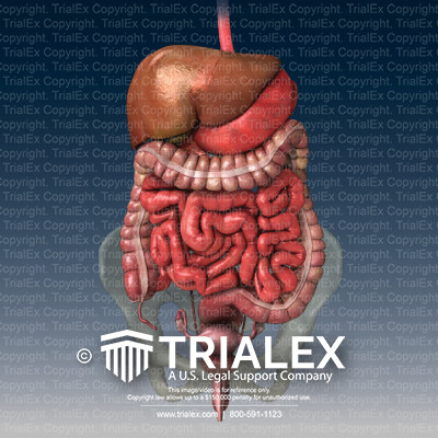
Normal Abdominal Organs Anatomy - TrialExhibits Inc. from cdn.trialexhibitsinc.com The abdomen contains all the digestive organs, including the stomach, small and large intestines, pancreas, liver, and gallbladder. Organs of abdomen diagram 1.5 / 10 (2 votes) organs of abdomen diagram in this image, you will find liver, stomach, pancreas, kidney, large intestine, gallbladder, small intestine, uterus in it. the abdomen consists of: A fold of peritoneum may completely cover certain organs, whereas it may cover only one side of organs that usually lie closer to the abdominal wall. Spleen is 1 inch thick, 3 inches broad and 5 inches long. It may include other soft tissues found here, such as the fibrous membrane. We're going to take apart a plastic anatomy model and see what we can find in the abdomen. • abdominal walls • abdominal cavity • abdominal viscera
The gastrointestinal tract is an organ system that enables us to ingest food, digest it, absorb it, and then expel the remaining waste as faeces.
A) describe the general location, function, and relationships among the following structures: 84 anatomical diagrams and histological images with over 300 labeled anatomical parts. Teachme anatomy part of the teachme series the medical information on this site is provided as an information resource only, and is not to be used or relied on for any diagnostic or treatment purposes. The abdominal cavity is the part of the body that houses the stomach, liver, pancreas, kidneys, gallbladder, spleen, and the large and small intestines.the diaphragm marks the top of the abdomen and the horizontal line at the level of the top of the pelvis marks the bottom. Finally, the abdomen contains an extensive membrane called the peritoneum. Labeled examination graphic with hypochondrium, hypogastrium and hypochondriac as. We'll identify as many organs as we can, see how they fit into the. The abdomen contains all the digestive organs, including the stomach, small and large intestines, pancreas, liver, and gallbladder. It is bounded superiorly by the xiphoid process and costal margins, posteriorly by the vertebral column and inferiorly by the pelvic bones and inguinal ligament. It may include other soft tissues found here, such as the fibrous membrane. The majority of these organs are encased in a protective membrane termed the peritoneum. For anatomy or health designs on white background for anatomy, medical, body, stomach, medicine, heart. The abdominal wall surrounds the abdominal cavity, providing it with flexible coverage and protecting the internal organs from damage.
The remainder of the abdominal organs are entirely covered with the visceral layer and are called intraperitoneal organs abdominal anatomy. It is bounded superiorly by the xiphoid process and costal margins, posteriorly by the vertebral column and inferiorly by the pelvic bones and inguinal ligament.